Meniscal Injuries - Overview

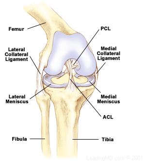
The medial meniscus and lateral meniscus are specialized structures within the knee. These crescent-shaped shock absorbers between the tibia and femur have an important role in the function and health of the knee. Once thought to be of little use, the menisci (plural) were routinely removed when torn.
Now we know that the menisci contribute to a healthy knee because they play important roles in joint stability, force transmission, and lubrication. When possible, they are repaired if injured. There are even experimental attempts to replace a damaged meniscus, possibly an important advance in orthopaedic medicine.
There are two categories of meniscal injuries - acute tears and degenerative tears.
- An acute tear usually occurs when the knee is bent and forcefully twisted, while the leg is in a weight bearing position. Statistics show that about 61 of 100,000 people experience an acute tear of the meniscus.
- Degenerative tears of the meniscus are more common in older people. Sixty percent of the population over the age of 65 probably has some sort of degenerative tear of the meniscus. As the meniscus ages, it weakens and becomes less elastic. Degenerative tears may result from minor events and there may or may not be any symptoms present.

What are the menisci?
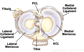
The two menisci of the knee are crescent-shaped wedges that fill the gap between the tibia and femur. The menisci provide joint stability by creating a cup for the femur to sit in. The outer edges are fairly thick while the inner surfaces are thin. If the menisci were missing, the curved femur would move on the flat tibia.
The medial meniscus, located on the inside of the knee, is more of an elongated "C"- shape, as the tibial surface is larger on that side. The medial meniscus is more commonly injured because it is firmly attached to the medial collateral ligament and joint capsule. The lateral meniscus, on the outside of the knee, is more circular in shape. The lateral meniscus is more mobile than the medial meniscus as there is no attachment to the lateral collateral ligament or joint capsule.

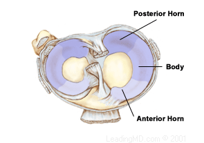
The outer edges of each meniscus attach to the tibia by the short coronary ligaments. Other short ligaments attach the ends of the menisci to the tibial surface. The inner edges are free to move because they are not attached to the bone. This lets the menisci change shape as the joint moves. The front portion of the meniscus is referred to as the anterior horn, the back portion is the posterior horn, and the middle section is the body.
Under the microscope, the meniscus is fibrocartilage that has strength and flexibility from collagen fiber. Its resilience is due to the high water content in the spaces between the cells. There is not much blood supply to the menisci. Blood flows only to the outer edges from small arteries around the joint. The poor blood supply to the inner portion of the meniscus makes it difficult for the meniscus to heal.

What does the meniscus do?
The meniscus acts as a shock absorber for the knee by spreading compression forces from the femur over a wider area on the tibia.
- The medial meniscus bears up to 50% of the load applied to the medial (inside) compartment of the knee.
- The lateral meniscus absorbs up to 80% of the load on the lateral (outside) compartment of the knee.
- During the various phases of the walking cycle, forces shift from one meniscus to the other, and forces on the knee can increase to 2 - 4 times body weight.
- While running, these forces on the knee increase up to to 6 - 8 times body weight. There are even higher forces when landing from a jump.
- The important role of the meniscus in force transmission can be seen when the menisci are removed.
- If the menisci are removed, the forces are no longer distributed over a wide area of the tibia. Without the medial meniscus, the tibial contact area is decreased 50 - 70%. This means the same forces from the femur are concentrated on a smaller area of the tibia.
- When the lateral meniscus is removed, there is a 45 - 50% decrease in contact area. This results in a 200 - 300% increase in contact pressure, which can eventually damage the cartilage on the ends of the bones. This can lead to degenerative arthritis.
- In the 1960s and 1970s, it was common to remove a damaged meniscus entirely. This frequently led to early degenerative arthritis in many patients.
- Removing the entire medial meniscus can lead to a bow-legged deformity and medial joint arthritis.
- Removing the entire lateral meniscus can cause a knock-kneed deformity and lateral joint arthritis.
What is a meniscus injury?
Patients describe meniscal tears in a variety of ways. Knowing where and how a meniscus was torn helps the doctor determine the best treatment.

- Location -A tear may be located in the anterior horn, body, or posterior horn. A posterior horn tear is the most common. The meniscus is broken down into the outer, middle, and inner thirds. The third in which the tear is located will determine the ability of the tear to heal, since blood supply in that area is critical to the healing process. Tears in the outer 1/3 have the best chance of healing.
- Pattern - Meniscal tears come in many shapes. The pattern of the tear influences the doctor's decision on treatment. Examples of the various patterns are:
A complex tear includes more than one pattern.
- Completeness - A tear is classified as being complete or incomplete. A tear is considered complete if it goes all the way through the meniscus and a piece of the tissue is separated from the rest of the meniscus. If the tear is still partly attached to the body of the meniscus, it is considered incomplete.
- Stability - A tear can be stable or unstable. A stable tear does not move and may heal on its own. An unstable tear allows the meniscus to move abnormally and is likely to be a problem if it is not surgically corrected.
Next